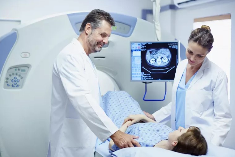
An X-ray or ultrasound test’s visual representation is insufficient for some medical conditions. More specific information is provided by CT scan images than by standard X-rays. So, to see your tissues, blood arteries, and bones in detail a Computed Tomography (CT) scan is used.
Through computer processing, a series of X-ray images collected from various angles around your body are combined in a CT scan. This would create a cross-sectional image, or slices, of your body’s soft tissues, blood arteries, and bones. Read below to know in detail about CT scan:
What are the uses of CT scan?
Here are some of the most common uses of ct scan:
- Structural and assessment of shape of body parts
- Diagnosis of injury or trauma
- Diagnosis of disease like cancer
- Diagnosis of vascular disease
- Help in planning for radiotherapy
- Help in planning particular surgeries
- Measurement of strength of bone
- Visual help in radiotherapy administration
- Visual help in certain interventional procedures such as needle aspiration or biopsy
- Alternative to some types of diagnostic or exploratory surgery
How do CT scans work?
The ct scan specifically use a narrow, circling X-ray beam on one area of your body. This is a collection of photos taken from various perspectives. This data is sent into a computer to produce a cross-sectional image. This two-dimensional (2D) scan displays a “slice” of your body’s inside, much like one piece in a loaf of bread.
To create several slices, this process is repeated. The computer builds a detailed picture of your organs, bones, or blood vessels by layering these scans on top of each other. A medical professional can use this kind of scan, to examine a tumor from all angles to get ready for surgery.
What can you expect during a CT scan?
Usually, during the test you will lie on a table, similar to a bed, on your back. A medical professional might give an intravenous (into your vein) contrast dye injection if your test needs it. You can experience flushing or taste something metallic after using this color.
When the scan starts: The bed enters the doughnut-shaped scanner gradually. You should now try to remain as still as possible because any movement will cause the images to get blurred. A brief hold of breath, usually lasting no more than 15 to 20 seconds, may also be required.
Your healthcare provider needs to see pictures of the area that the scanner captures. A CT scan is quieter than an MRI (magnetic resonance imaging) scan. After completion of the test, the table retracts from the scanner.
After the CT scan
While the radiographer analyzes the images, you might be requested to wait. Sometimes it is necessary to take extra pictures. When the radiographer says the process is complete, you can get dressed and leave after they have taken enough clear pictures.
If you were given the iodinated contrast material, you may have to spend a brief amount of time in the department following the procedure. The radiology specialist with medical training evaluates the scans. You will need to schedule a follow-up session because the results normally go to the specialist who referred you.
Final thoughts
If your healthcare provider suggests a CT scan, it is normal to be skeptical or perhaps a little concerned. However, CT scans are a low-risk, painless procedure that can assist medical professionals in identifying a variety of illnesses. Getting a precise diagnosis helps your medical professional in deciding on the most appropriate diagnosis.

